Taser Stratbucker Safety Studies 1996
Download original document:

Document text

Document text
This text is machine-read, and may contain errors. Check the original document to verify accuracy.
SECTION VII: p. 1

A1""BU' {'"'KE~P

u~ll""P
_,-1"i

•
•

~___,-

& Associates
10744 West Center Road * Omaha, Nebraska 68124' (402) 399-9500' fax (4.Q2) 571-4017

INTRODUCTION
I

In late 1995, I was contacted by representatives of AIR TASER, Incorporated seeking to perfonn
tl
safety studies of the impulse generator module of the AIR TASER product. The goal of the
study was to perform an analysis to establish a margin ofsafety for the AIR TASER Model
#34000 by testing significant increases in relevant electrical characteristics and evaluating the
physiological response.

I
I

PROCEDURE
Over the past decade, I have tested a number of similar devices both physically and
physiologically in my laboratory and have become quite familiar with the necessary procedures
to accurately accommodate such testing. Accordingly, I agreed to commence such testing upon
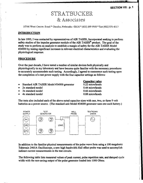
the completion of a test power supply with the four capacitor settings as follows:

•
•
•
•

Standard AIR TASER Model #34000 generator
2x standard model
3x standard model
4x standard model

Capacitor value
0.22 microfarads
0.44 microfarads
0.66 microfarads
0.88 microfarads

The tests also included each ofthe above noted capacitor sizes with one, two, or three 9 volt
batteries as a power source. (The standard unit Model #34000 generator uses one such battery.)
POWER
SUPPLY

(0
•

8
8

TEST
GENERATOR

®
jJF

@
jJF

HALL
PROBE

~

TEKTRONIX

2465A

E9

LOAD

@
jJF

aD
jJF

In addition to the familiar physical measurements of the pulse wave fonn using a 100 megahertz
Tektronix 2465A Oscilloscope, a new high bandwidth Hall effect probe was used to accomplish
indirect current measurements in the test circuits.
The following table lists measured values ofpeak current, pulse repetition rate, and damped cycle
width with the non-arcing output of the pulse generator loaded into 1000 Ohms.

SECTION VII: p. 2
•

Electrical Output Measurements
CapacitaDce
(MIcrofarads)

Battery IDput
(Volts)

Peak CurreDt
(Amperes)

IDterval
(MIDlsecoDds)

Pulse Wldtb
(MIcrosecoDds)

Load
(Obms)

0.11
0.44
0.66
0.88

9
9
9
9

10
13
16
18

130
600
750
1000

65

1000
1000
1000
1000

0.11
0.44
0.66
0.88

18
18
18
18

9.1
14
16
18

80
150
350
500

6.9
9.5
11.4

0.12
0.44
0.66
0.88

17
17
17
17

8

44
88
160
400

7
10

1Z

15
17

9.4
11
11

1Z

11

13

1000
1000
1000
1000
1000
1000
1000
1000

Most importantly, the above described protocol was to be evaluated in anaesthetized animals of
representative size and cardiac status to adult humans. Accordingly, such physiologic testing was
performed using market sized farm swine conveniently available to the laboratory.

On January 11, 1996 an animal test was performed using the identical protocol outlined in the
physical study. An 18.2 kg Hampshire shoat, the standard subject used in many cardiac safety
studies, was pre-medicated with atropine sulfate (0.02 mg/kg) intramuscularly. Shortly
thereafter, Ketamine (lOmg/kg) mixed with Xylazine (2.01mg/kg) were given intramuscularly in
serial doses spaced by 15-20 minutes to affect stage I to stage II anesthesia for the one hour
duration of the procedure. The airway was carefully managed, but intubation was not required
nor was assisted ventilation. At the conclusion of the procedure, the animal was allowed to
recover and was returned to its pen in excellent condition.
In each of the twelve steps in the 4 x 3 protocol described above, the animal was stimulated with
the device via output electrodes placed on the left hindquarter to determine skeletal muscle
response, vertically oriented on the anterior abdomen at the umbilicus to asses mid-abdominal
response and fmally with both vertical and transverse orientation at the level of the cardiac apex
to assess any possible affect on cardiac rhythm. In this latter regard, it should be noted that a
three channel battery powered cardiograph unit was continuously employed to accomplish
orthogonal lead axes. Such technique overcomes the serious deficiencies ofseveral prior reports
in which the pulse generator axis coincides with a non-dominant electrocardiographic axis of the
heart, nearly obliterating the animal's electrocardiogram and erroneously raising doubt as to the
expected immunity of the cardiac rhythm to the effects of body surface electric discharges. 1

I, !

SECTION VII: p. 3

•
I

!

I

I
I
I

RESULTS

Of the more than 48 discharges of five seconds duration, there was no case in which the animal
revealed any cardiac ectopy or myocardial injury. The cardiac tissue proved resistant to
stimulation despite progressively increased skeletal muscle effects noted as the storage
cllpacitors and the battery output were increased by several hundredpercent. Respiration was
briefly arrested during the application ofsome of the chest discharges, but returned
2
spontaneously upon cessation ofstimulation. Several other mild autonomic effects such as
increased heart rate and respiration rate were observed with the higher potency discharges. Both
respiration and heart rate returned to normal in a matter ofa few minutes. On the day following
this rigorous protocol, the animal appeared to be completely normal with the exception of a few
lingering electrical "signature" marks on its chest and abdomen.

DISCUSSION
3

These experiments corroborate our earlier findings in consulting reports and peer review
journals that the electrical emissions from stun type pulse generators, delivered to the body
surface in the recommended manner do not cause serious cardiac rhythm abnormalities in the
otherwise healthy adult heart. As this study investigated electrical outputs equivalent to 400%
the capacitance and 300% the battery voltage of the standard AIR TASER Model #34000, an
adequate margin of safety4 appears to exist.

Respectfully Submitted,

Robert A. Stratbucker, MD, Ph.D.

-

O.Z. Roy and A.s. Podgorski, Tests on a Shocking Device - the Stun Gun. Med. & BioI. Eng. & Compu!., 1989,
27, 445-448.
, Note: The AIR TASER was designed with a pre-programmed timing cycle in light of the potential for respiratory
interruption. The unit automatically provides four I second pauses during each 30 second discharge to allow the
aubjectto breathe. During this study, the animal promptly resumed normal breathing upon cessation of electrical
stimulus. Hence, the four I second breaks allow the target to take four full breaths every 30 seconds, minimizing
the risk of anoxia. (However, interruption of respiration for a full 30 seconds poses little health risk.)
, Robert A. Slratbucker and Matthew G. Marsh. IEEE The Relative Immunity of the Skin and Cardiovascular
System to the Direct Effects of High Voltage - High Frequency Component Electrical Pulses. Proc. IEEE
Engineering in Medicine & Biology Conference, October 1993, San Diego, CA.
• Pearce, J.A. et al: Myocardial Stimulation with Ultrashort Duration Current Pulses. PACE, Vol. 5, JanuaryFebruary 1982.
I

